
Tips Memilih Mikroskop Fluoresensi yang Tepat: Widefield, Inverted, Confocal, atau Live-Cell?
A. Mengenal Mikroskop Fluoresensi
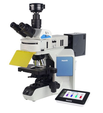
Gambar 1. MShot Fluorescence Microscope
Mikroskop fluoresensi merupakan alat utama yang berfungsi untuk mengamati fisiologi sel. Teknik mikroskop fluoresensi digunakan untuk mengamati zat-zat fluoresen di bawah mikroskop. Keunggulan dari teknik ini dibanding mikroskop lainnya, yaitu memiliki sensitivitas dan spesifisitas yang tinggi.
Spesimen yang dideteksi menggunakan mikroskop fluoresensi disinari dengan cahaya yang memiliki panjang gelombang relatif pendek, biasanya biru atau ultraviolet (UV). Pengamatan spesimen melalui filter penghalang yang menyerap cahaya yang panjang gelombangnya pendek untuk digunakan sebagai penerangan dan meneruskan fluoresensi, sehingga hasil terlihat terang dengan latar belakang yang gelap.
Proses dasar fluoresensi meliputi penyerapan energi cahaya (foton) oleh indikator, yang diikuti oleh emisi sebagian energi cahaya tersebut (sebagian foton lain) beberapa nanosekon kemudian. Karena sebagian energi hilang dalam proses ini, foton yang dipancarkan memiliki energi yang lebih rendah daripada foton yang diserap. Cahaya dengan panjang gelombang pendek (biru) memiliki energi yang lebih tinggi dibandingkan cahaya dengan panjang gelombang panjang (merah). Oleh sebab itu, cahaya yang dipancarkan dari suatu indikator biasanya memiliki panjang gelombang yang lebih panjang daripada cahaya yang diserap (eksitasi). Pemisahan cahaya tersebut umumnya dilakukan dengan filter optik dan keberhasilan pencitraan bergantung pada ketepatan pemilihan filter yang sesuai dengan indikator yang digunakan.
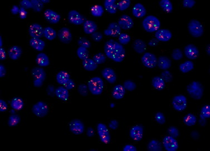
Gambar 2. Hasil Pengamatan menggunakan MShot Fluorescence Microscope
B. Fluoresensi Tradisional (Widefield Fluorescence Microscopy)
Widefield Fluorescence Microscopy secara sederhana memiliki alur kerja dimana sinar cahaya paralel secara bersamaan menerangi seluruh spesimen (bidang pandang yang luas) untuk mengeksitasi (melalui blok filter) fluorofor yang ada didalamnya. Secara tradisional, cahaya eksitasi disuplai oleh lampu merkuri atau xenon bertekanan tinggi. Adapun panjang gelombang yang diperlukan dipilih dengan filter optik khusus. Namun, semakin berkembangnya teknologi bentuk cahaya eksitasi dapat menggunakan LED yang terang dan berpanjang gelombang tunggal.
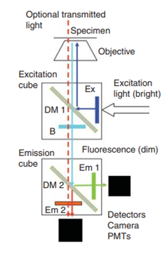
Gambar 3. Jalur Cahaya Dasar pada Mikroskop Fluoresensi
Sebuah kubus filter eksitasi yang berisi cermin dikroik (DM1) mengarahkan cahaya eksitasi (dari laser yang telah di filter dengan filter eksitasi) ke spesimen dan meneruskan fluoresensi yang dipancarkan ke kubus emisi untuk pemisahan lebih lanjut. Filter penghalang B mencegah cahaya eksitasi mencapai detektor. Fluoresensi yang dipancarkan dipisahkan oleh DM2 menjadi 2 sinar. Filter emisi (Em 1 dan Em 2) memblokir cahaya yang tidak diinginkan. Fluoresensi dideteksi oleh kamera (wide field) atau PMTs (pemindaian laser). Iluminasi transmisi pada gambar bidang terang, cahaya dengan panjang gelombang panjang yang dipilih.
Keunggulan:
- Seluruh bagian spesimen dapat diamati secara bersamaan dan gambar mudah direkam menggunakan kamera.
- Menghasilkan gambar yang bagus untuk spesimen berupa lapisan sel tunggal dan organel yang sangat tipis menempel erat di permukaan kaca.
- Pengambilan gambar yang lebih cepat.
- Biaya rendah, metodenya sederhana, dan sistemnya yang fleksibel.
Kekurangan:
- Menghasilkan gambar dengan kontras dan resolusi spasial yang rendah karena difraksi yang dibatasi dan proyeksi cahaya yang tidak fokus ke bidang gambar tunggal.
- Menghasilkan gambar yang tumpang tindih karena difraksi optik yang menyebabkan ukuran objek lebih besar dari objek aslinya. Hal ini membuat gambar yang dihasilkan akan buram. Sehingga, perlu adanya pemeriksaan pada sampel bahan fluoresen yang jaraknya lebih jarang.
- Berpotensi terjadinya artefak bayangan akibat pencahayaan tidak merata.
- Perlu penyelarasan berbagai kamera untuk memastikan registrasi pixel saat menggunakan beberapa indikator.
Berikut merupakan produk Fluorescence Microscope dari MSHOT yang dapat Anda pesan melalui PT. Indogen Intertama sesuai dengan kebutuhan lab Anda.
Tabel 1. Produk Fluorescence Microscope MSHOT
| Nomor Katalog | Deskripsi Produk |
| ML45-FL | 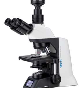
Features: ● Infinity-corrected optical system with flat-field objectives ● Coded objective turret for intelligent selection ● Intelligent digital control and user-friendly operation ● Universal and versatile application scenarios ● Cost-effective, high-performance solution
Specifications: ● Main Body: Infinity-corrected optical system; built-in ECO standby (auto-off when idle), Type-C control, USB/DC modular power supply, and adjustable color temperature. ● Eyepiece Tube: 30° tilting trinocular head, 55/45 split, 30° inclined, interpupillary adjustment range: 50–75 mm; 3-position trinocular head (optional). ● Eyepiece: Widefield WF10X / F.N.23. ● Objective Lenses: A-Plan plan achromatic objectives: 4×/10×/40×/100× (standard), 20× (optional). Optional accessories include phase contrast objectives, fluorescence objectives, semi-apochromatic objectives, etc. ● Focusing Mechanism: Coaxial coarse/fine focusing mechanism; focusing range: 25 mm; fine adjustment resolution: 0.002 mm. ● Nosepiece: Inward 5-position coded nosepiece with brightness memory function. ● Stage: Stage size: 232 × 140 mm; travel range: 76 × 50 mm. ● Condenser: Abbe condenser, NA 1.1. ● Transmitted Illumination System: 3W LED light source with continuously adjustable color temperature. ● Epi-fluorescence Module: Standard upright triple-color digital fluorescence module with 3 channels (slider switch); optional 6-position motorized turret module with 3 standard channels. Standard filter sets: U (EX: 360–390 nm; DM: 415 nm; EM: 460/50 nm), B (EX: 470/40 nm; DM: 505 nm; EM: 535/40 nm), G (EX: 510–550 nm; DM: 565 nm; EM: 605/55 nm). Custom fluorescence wavelengths available on request. ● Digital Display: Real-time display of objective magnification, brightness, broadcast status, ECO mode, and other related information. ● Intelligent System: Built-in ECO auto-off function; optional voice announcement.
|
| MF31 | 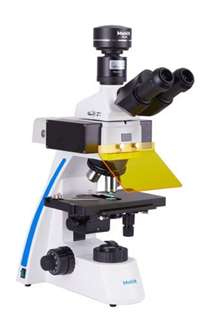
Features: ● Infinity plan achromatic objective system ● Wide-field eyepiece for binocular or trinocular observation LED fluorescence excitation module ● Brightfield and fluorescence dual-mode observation ● Excellent accessory compatibility
Specifications: ● Eyepiece: Wide field eyepiece WF10X/22mm ● Viewing Head: Seidentopf trinocular head, 30° inclined ● Objective: ➔ Infinity plan achromatic objective 4X ➔ Infinity plan achromatic objective 10X ➔ Infinity plan achromatic objective 40X ➔ Infinity plan achromatic objective 100X ● Fluorescence Module: ➔ Blue (B) excitation: 460–490nm ➔ Green (G) excitation: 510–550nm (Optional) ➔ Ultraviolet (UV) excitation: 330–380nm (Optional) ➔ Blue-Violet (BU) excitation: U: 330–380nm; B: 460–490nm (Optional) ➔ Blue-Green-Ultraviolet (BGU) excitation: U: 330–380nm; B: 460–490nm; G: 510–550nm (Optional) ● Focusing System: Coaxial coarse and fine focusing system, with tension adjustment and limit stopper, fine focusing graduation: 2μm, coarse stroke per rotation: 40mm, fine stroke per rotation: 0.2mm, focusing range: 24mm ● Nosepiece: Inward quadruple or quintuple revolving nosepiece (Optional) ● Stage: Double-layer mechanical stage (Size: 210mm × 140mm, movement range: 76mm × 50mm) ● Transmitted Illumination: ➔ Abbe condenser N.A. 1.25 ➔ White LED, adjustable brightness ● Camera Adapter: 1X C-mount (0.5X C-mount Optional) ● Power Cord: Standard 3-pin power cord ● Dust Cover: Dedicated dust cover
|
| MF23 | 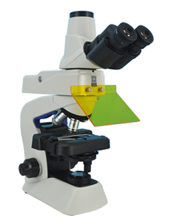
Features: ● LED fluorescence excitation ● Infinity-corrected optical system ● Brightfield and fluorescence observation ● High transmission objectives ● Accessory compatibility
Specifications: ● Optical system: Infinity optical system ● Eyepiece: Wide field WF10X/FN.20mm (anti-fungus) ● Observation head: Trinocular head with triple light split 0 / 50% /100% ● Objectives: ➔ Infinity plan achromat 4X/0.10, working distance 27.8mm ➔ Infinity plan achromat 10X/0.25,working distance 8.0mm ➔ Infinity plan achromat 40X/0.65,working distance 0.60mm ➔ Infinity plan achromat 100X/1.25(spring), working distance 0.13mm ● Focusing system: Fine and coarse adjustment with tension and limited stopper, minimum adjustment gradations:2.5um,coarse movement stroke:15mm ● Nosepiece: Quadruple fixed nosepiece ● Stage: ➔ Wire movement mechanical fixed stage, size 120mmx132mm, travelling range 76mmx30mm ➔ Single specimen holder ● Condenser: Abbe condenser, NA 1.25 (with immersion oil), inter set aperture diaphragm ● Fluorescence illumination: ➔ LED fluorescence illuminator ➔ 3W LED ➔ Blue excitation: EX460~490nm, DM>500nm, EM>510nm ● Transmitted illumination: ➔ Built-in transmitted illumination system, LED power Consumption 0.5W(nominal values) ➔ Optional: 6V 20W halogen lamp ● Power source: AC, 100-240V, 50~60Hz
|
| MF43-N | 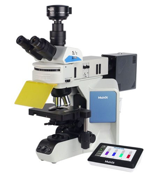
Features: ● LED fluorescence excitation ● Infinity-corrected optical system ● Brightfield and fluorescence observation ● High transmission objectives ● Accessory compatibility
Specifications: ● Eyepiece: Wide field 10X/23mm; 10X/25mm (Optional) ● Viewing Head: ➔ Seidentopf trinocular viewing head, high eyepoint, 30° inclined, interpupillary distance: 50–75mm ➔ Visual:Digital light split ratio = 100:0 or 20:80 or 0:100 ● Objective: ➔ Plan semi-apochromatic objective 4X / 10X / 40X / 100X ➔ Plan semi-apochromatic objective 20X (Optional) ● Epi-Fluorescence Illumination System: ➔ 4-channel light source MG-120 / High-efficiency fluorescence light source MG-150 (Optional) ➔ Touch screen controller ➔ Empty epi-fluorescence illuminator FL-43 (6-position turret, standard with BGU channels, optional YVR, etc.) ➔ FB-U-M band EX: 375/28nm; DM: 415nm; EM: 460/50nm ➔ FB-B-M band EX: 470/40nm; DM: 505nm; EM: 530/40nm ➔ FB-G-M band EX: 530/40nm; DM: 565nm; EM: 605/55nm ➔ V band EX: 405/30; DM: 440; EM: 450LP (Optional) ➔ R band EX: 620/50; DM: 655; EM: 692/45 (Optional) ➔ Y band EX: 560/40nm; DM: 600nm; EM: 610nmLP (Optional) ➔ Dual-band B/G EX: 470/40 & 575/35; EM: 525/40 & 625/60 (Optional) ➔ Royal blue B2 EX: 450/50; DM: 485; EM: 495LP (Optional) ➔ Yellow G2 EX: 560/40; DM: 600; EM: 610LP (Optional) ● Focusing System: Coaxial coarse and fine focusing system, focusing stroke: 25mm, with coarse upper limit stopper, coarse tension adjustable, fine focusing graduation: 1μm ● Nosepiece: Inward coded quintuple revolving nosepiece ● Stage: Movement range: 80×50 mm, precision: 0.1mm ● Transmitted Illumination: ➔ Warm white LED light source, continuously adjustable brightness, equipped with intensity manager ➔ Abbe condenser N.A. 1.1, Köhler illumination
|
C. Inverted Fluorescence Microscope
Sistem optik Inverted Fluorescence Microscope sama seperti Wide Field Fluorescence Microscope. Perbedaan mendasarnya adalah lensa objektif memproyeksikan gambar sampel dari bawah. Inverted microscope sangat sesuai untuk sel yang dikultur pada coverslip-bottom dish atau multiwell plate.
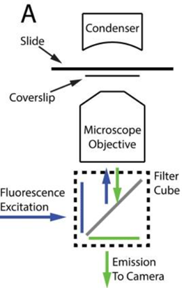
Gambar 4. Skema Teknik Inverted Fluorescence Microscope
Sampel diletakkan di antara kaca objek dan kaca penutup. Lensa kondensor menyalurkan cahaya untuk mengamati cahaya yang ditransmisikan melalui sampel. Lensa objektif mengumpulkan cahaya dari sampel dan menyalurkan cahaya eksitasi untuk mikroskop fluoresensi. Kubus filter terdiri dari filter eksitasi (biru), filter emisi (hijau), dan cermin dikroik (abu-abu). Filter eksitasi dan emisi masing-masing memilih panjang gelombang yang akan menerangi sampel dan direkam oleh kamera. Adapun cermin dikroik memantulkan cahaya eksitasi ke sampel sekaligus meneruskan cahaya emisi ke kamera.
| Nomor Katalog | Deskripsi Produk |
| MIX60-FLD | 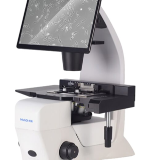
Features: ● Integrated tablet display for instant, standalone imaging ● Infinity-corrected inverted optical system ● Motorized fluorescence turret with 5 standard color channels ● One-click acquisition and intuitive software interface ● Space-saving all-in-one design for busy laboratories
Specifications: ● Main Unit: Infinite-correction optical system ● Stage: ➔ Fixed stage ➔ Stage extension board ➔ XY-axis stage movable ruler ➔ Universal sample holder for various containers: Round transparent drip plate: Φ118 mm ● Condenser: ➔ Removable condenser, N.A.: 0.3; WD: 72 mm ➔ Suitable for objectives: 4×, 10×, 20×, 40×, 60× ● Phase Ring Plate: One 10× phase ring; one shared phase ring for 20×/40× ● Bright-field Illumination: ➔ 3 W full spectrum white LED, continuously adjustable brightness ➔ Detachable filter mount for 34 mm filters ➔ Aperture diaphragm with manual open/close ● Nosepiece: 5-hole motorized coded nosepiece, software-controlled switching ● Objectives: Standard: FL 4×; FL Ph 10×; FL Ph 20×; FL Ph 40×; other magnifications & types optional ● Fluorescence Illumination: Motorized 6-position fluorescence turntable; ● Focusing Mechanism: Coaxial coarse/fine focusing with limit & lock; Low-position coaxial knobs; fine focus division: 2 μm ● Imaging System: ➔ 1/1.6-inch color CMOS ➔ 12.5 MP, 2.0 μm × 2.0 μm ➔ Frame rate: 28 fps ➔ 15V 4A
|
| MIX60-FL | 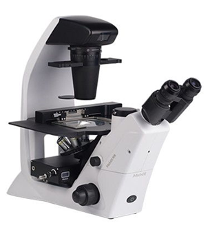
Features: ● Inverted optical configuration for samples in vessels ● Infinity-corrected optics ● Phase contrast or fluorescence options ● Long working distance objectives ● Stable, ergonomic design
Specifications: ● Host: 200mm infinity optical system, the body is equipped with ECO light-off function and can draw power ● Eyepiece: ➔ WF10X/22 plan eyepiece, high eye point, adjustable diopter ➔ WF10X/23 plan eyepiece, high eye point, adjustable diopter ➔ Centering telescope ● Eyepiece tube: Inclination angle 45°, interpupillary distance 50-70mm, body split ratio = 100:0 or 0:100 ● Nosepiece: Quintuple nosepiece, ball bearing with anti fungus device ● Stage: ➔ Fixed stage size: 267.4mm×271.4mm ➔ XY stage moving ruler, mechanical movement, movement range: 110mm×75mm ➔ With stage expansion board ● Universal sample holder: Match slides and different petri dish sizes ● Drip board: Round transparent drip board: φ118mm ● Condenser: ➔ Numerical aperture N.A.: 0.3, working distance WD: 72mm ➔ Compatible Objective Magnifications 4X, 10X, 20X, 40X, 60X ➔ Detachable condenser, container height ≥19cm can be placed on the stage ● Phase ring plate: 10X ring, 20X/40X ring ● Focusing mechanism: Coaxial coarse/fine focus, with tension adjustable and up stop, minimum division of fine focusing is 2μm. ● Transmitted illumination system: ➔ 3W full spectrum white LED, brightness continuously adjustable ➔ The filter holder can be inserted into φ34mm color filters (blue, green, yellow, etc.) and is removable ➔ With aperture diaphragm, aperture blades, manual control switch Digital display screen ➔ Displays the current working status, including ECO, voice function, objective lens multiple and brightness, etc., and can perform voice broadcast of the current objective lens multiple. ● Objective lens: ➔ Semi-apo M-UPLFN objective 4X ➔ Long working distance fluor objective LPlan FL 40X ➔ Semi-apo fluor-phase contrast objective LPlan FLPH 10X ➔ Semi-apo fluor-phase contrast objective LPlan FLPH 20X ● Fluorescence module: ➔ MIF2-UBG:B:450-490nm, G:510-550nm, U:360-390nm ➔ MIF2-UBY:B:450-490nm, Y:540-580nm, U:360-390nm
|
| MF52-N | 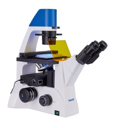
Features: ● Inverted optical configuration for samples in vessels ● Infinity-corrected optics ● Phase contrast or fluorescence options ● Long working distance objectives ● Stable, ergonomic design
Specifications: ● Eyepiece: ➔ Wide field SWF10X, FN 22mm, high eyepoint, one diopter is adjustable ➔ Centering telescope ● Observation tube: 45° inclined, interpupillary distance 53–75mm ● Nosepiece: Quintuple nosepiece, ball bearing with anti fungus device ● Objectives: ➔ Long working distance plan objective lens M-UPLFLN4X/0.13 Working distance: 17.15mm ➔ Long working distance plan objective lens Plan 40X/0.58 Working distance: 2.5mm ➔ Long working distance plan phase contrast objective lens Plan10X/0.26 PH working distance: 10.5mm ➔ Long working distance plan phase contrast objective Plan20X/0.45 PH Working distance: 5mm ➔ Long working distance plan objective Plan10X/0.25, working distance: 4.1mm(Optional) ➔ Long working distance plan objective lens Plan20X/0.45 Working distance: 5mm(Optional) ➔ Long working distance plan objective Plan40X/0.58, working distance: 2.5mm(Optional) ➔ Long working distance plan phase contrast objective 4X(PH)(Optional) ➔ Long working distance semi-apochromatic plan objective 4X/10X/20X/40X(FL)(Optional) ➔ Long working distance 10X semi-apochromatic plan phase contrast objective 10X/20X(FLPH)(Optional) ● LED fluorescence attachment and illuminating system: ➔ MIF-BGU-BP — B:450–490nm, G:510–550nm, U:361–389nm ➔ MIF-UBY-BP — U:360–390nm, B:460–495nm, Y:540–580nm(Optional) ➔ MIF-BGU-LP — B:460–495nm, G:510–550nm, U:330–385nm(Optional) ➔ MIF-BG-BP — B:460–495nm, G:528–553nm(Optional) ● Phase contrast ring plate: 10X ring, 20X/40X ring ● Focus system: Coaxial coarse/fine focus, with tension adjustable and up stop, minimum division of fine focusing is 2μm ● Stage: ➔ Fixed stage overall size is 227mm×208mm ➔ Mechanical moving 135mm×77mm ➔ Transparent glass round stage overall size is Φ118mm, inner Φ68mm ➔ Petri dish holder 1 — Inside locating slot size: 86mm (W)×129.5mm (L) (suitable for circular culture dish Φ90mm) ➔ Petri dish holder 2 — Inside locating slot size: 34mm (W)×77.5mm (L) (suitable for circular culture dish Φ68.5mm) ➔ Petri dish holder 3 — Inside locating slot size: 57mm (W)×82mm (L) (suitable for circular culture dish Φ60mm) ➔ Petri dish holder 4 — Inside locating slot size: 29mm (W)×77.5mm (L) (suitable for circular culture dish Φ35mm) ● Transmitted illumination system: ➔ White LED lamp with brightness adjustable ➔ Push-pull type condenser, working distance 55mm ➔ Green filter ● Camera adapter: Internal set 0.75X C-mount ● Accessories: ➔ Allen driver ➔ Dust cover ➔ Fluorescence light shield
|
| MF53-N | 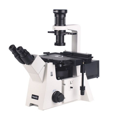
Features: ● Inverted optical configuration for samples in vessels ● Infinity-corrected optics ● Phase contrast or fluorescence options ● Long working distance objectives ● Stable, ergonomic design
Specifications: ● Eyepiece: ➔ Wide field Plan eyepiece WF10X/23mm, high eye point ➔ Centering telescope ● Observation tube: Binocular observation tube, 45° inclined, pupil distance adjust range 50–75mm, diopter adjustable ● Nosepiece: Quintuple revolving nosepiece with bearing inner location and anti-fungus device ● Objectives: ➔ Infinity long working distance plan achromatic objective Plan 4X/0.12, WD:10.8mm ➔ Long working distance semi-apochromatic fluorescence objective L Plan FL 10X/0.32, WD:7.1mm ➔ Long working distance semi-apochromatic fluorescence objective LWD Plan Fluor 40X/0.65, WD:3.0mm ➔ Infinity long working distance plan achromatic objective Plan 10X/0.26 PH, WD:10.5mm ➔ Infinity long working distance plan achromatic objective Plan 20X/0.45 PH, WD:5mm ➔ Infinity long working distance plan achromatic objective LWD Plan 4X/0.1, WD:11.98mm ➔ Infinity long working distance plan achromatic objective Plan 10X/0.26, WD:10.5mm ➔ Infinity long working distance plan achromatic objective Plan 20X/0.45, WD:5mm ➔ Infinity long working distance plan achromatic objective Plan 40X/0.58, WD:2.5mm ➔ Infinity long working distance plan achromatic objective Plan 10X/0.25 PH, WD:9.3mm ➔ Infinity long working distance plan achromatic objective Plan 20X/0.45 PH, WD:5mm ➔ Infinity long working distance plan achromatic objective Plan 40X/0.58 PH, WD:2.5mm ➔ Infinity long working distance semi-apochromatic fluorescence objective Plan Fluor 4X/0.13, WD:18.5mm ➔ Infinity long working distance semi-apochromatic fluorescence objective Plan Fluor 10X/0.3, WD:7.1mm ➔ Infinity long working distance semi-apochromatic fluorescence objective Plan Fluor 20X/0.45, WD:5.0mm ➔ Infinity long working distance semi-apochromatic fluorescence objective L Plan FL 40X/0.65, WD:3.0mm ➔ Infinity long working distance semi-apochromatic fluorescence objective Plan Fluor 60X ➔ Long working distance semi-apochromatic phase contrast objective 10X(FLPH) ➔ Long working distance semi-apochromatic phase contrast objective 20X(FLPH) ➔ Long working distance fluorescence phase contrast objective 40X(FLPH/1.0) ● LED fluorescence attachment and illuminating system: ➔ Individual channels LED light source MG120 ➔ 100W Broad-spectrum LED light source ➔ Sixtuple filter cube positions fluorescence attachment with 3 filter cubes, available to add and exchange filters ➔ Filter cube — Filters: ➔ U — EX: 375/28nm; DM: 415nm; EM: 460/50nm ➔ B — EX: 470/40nm; DM: 505nm; EM: 535/40nm ➔ G — EX: 530/40nm; DM: 565nm; EM: 605/55nm ● Sextuple fluorescence module and its filter cubes: filter cubes: ➔ FB-U-M — EX: 375/28nm; DM: 415nm; EM: 460/50nm ➔ FB-B-M — EX: 475/30nm; DM: 505nm; EM: 530/40nm ➔ FB-G-M — EX: 540/25nm; DM: 565nm; EM: 605/55nm ➔ FL-U-M — EX: 355/50nm; DM: 410nm; EM: 420nmLP ➔ FL-B-M — EX: 475/30nm; DM: 505nm; EM: 510nmLP ➔ FL-G-M — EX: 550/40nm; DM: 570nm; EM: 575nmLP ➔ FL-Y-M — EX: 560/40nm; DM: 600nm; EM: 610nmLP ➔ FB-Y-M — EX: 560/40nm; DM: 600nm; EM: 635/60nm ➔ FB-R-M — EX: 620/50nm; DM: 655nm; EM: 690/50nm ● Quintuple fluorescence module and its filter cubes: ➔ UV — EX: 355/50nm; DM: 400nm; EM: 415nmLP ➔ B — EX: 475/30nm; DM: 500nm; EM: 515nmLP ➔ G — EX: 530/40nm; DM: 570nm; EM: 590nmLP ➔ FB2-U-M — EX: 375/30nm; DM: 415nm; EM: 460/50nm ➔ FB2-B-M — EX: 475/30nm; DM: 505nm; EM: 530/40nm ➔ FB2-G-M — EX: 540/25nm; DM: 565nm; EM: 605/55nm ➔ FB2-Y-M — EX: 560/40nm; DM: 600nm; EM: 635/60nm ➔ FB2-R-M — EX: 620/50nm; DM: 655nm; EM: 690/50nm ➔ FL2-Y-M — EX: 560/40nm; DM: 600nm; EM: 610nmLP ● Focus system: Coaxial coarse and fine with limit and locking devices, low coaxial focus adjusting handle, Minimum adjustment graduation: 1μm ● Stage: Fixed stage 240mm×260mm, with low-positioned X/Y coaxial control knob, travel distance 135mm×85mm ● Transmitted illumination system: ➔ Long working distance condenser 72mm, NA 0.30 with triple phase contrast slider 10X/20X/40X ➔ LED brightness continuously adjustment ➔ Black color light shield ● Slide glass holder: ➔ Multi-function slide glass holder (Ø35mm) ➔ Multi-function slide glass holder (Ø90mm) ● Camera adapter: External 1X C-mount adapter |
D. Mikroskop Fluoresensi Konfokal (Confocal Fluorescence Microscopy)
Mikroskop Fluoresensi Konfokal menjadi solusi untuk peneliti yang terhambat dalam penggunaan Mikroskop Fluoresensi Tradisional. Laser-Scanning Confocal Microscope (LSCM) mampu menangkap pencitraan dengan lebih baik. Seluruh konsep utama eksitasi dan emisi fluoresensi berlaku di LSCM. Perbedaan utama terdapat di sumber eksitasi (laser), metode pemindaian berurutan dari sumber penerangan titik tersebut, serta deteksi intensitas cahaya yang dipancarkan dengan tabung photomultiplier (PMT).
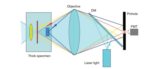
Gambar 5. Prinsip Kerja Mikroskop Konfokal Pemindaian Laser
Tujuan mikroskop konfokal untuk menyaring cahaya yang tidak fokus dari gambar. Hasil ini karena dalam mikroskop konfokal menggunakan lubang jarum yang dapat memastikan cahaya yang mencapai detektor hanya berasal dari titik yang setara (konfokal) pada spesimen tempat cahaya eksitasi difokuskan. Pemindaian titik eksitasi dan deteksi konfokal yang melintasi spesimen akan menghasilkan gambar yang tersusun berurutan piksel demi piksel, dengan menekan intensitas fluoresensi pada setiap posisi. Adapun sepasang cermin berosilasi yang saling tegak lurus sebagai pemindah lokasi titik fokus melintasi spesimen.
E. Sistem Live-Cell Imaging
Live-Cell Imaging System menggabungkan antara mikroskop, kontrol lingkungan, tahap otomatisasi, pengambilan gambar, dan analisis gambar dalam satu sistem. Sehingga, pengamatan sel dapat diamati dalam kondisi yang mendekati kondisi fisiologis selama riset berlangsung. Penggunaan Live-Cell Imaging System memungkinkan pengamatan proses biologis secara real-time. Kontrol lingkungan juga menjadi komponen penting seperti temperatur, CO2, kelembaban, dan kondisi media selama proses imaging. Adapun otomatisasi juga diperlukan untuk memungkinkan perpindahan sistem secara otomatis dari satu well ke well lainnya. Teknologi baru ini akan mempermudah peneliti khususnya yang berfokus pada penelitian sel.
Saat ini PT. Indogen Intertama menawarkan produk terbaru dari MSHOT berupa Live Cell Imaging System. Berikut daftar produk secara lengkap dapat dibaca pada Tabel 4.
Tabel 4. Produk Live Cell Imaging System MSHOT
| Nomor Katalog | Deskripsi |
| MCS31 | 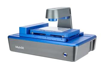
Features: ● Works inside standard incubators for stable, long-term time-lapse imaging. ● Dual phase-contrast and fluorescence observation modes. ● Supports culture flasks, dishes and multi-well plates (6-well to 384-well). ● Compact, easy-to-use design with repeatable positioning. ● Automated image capture for continuous live cell experiments.
Specifications: ● Brightfield Observation: ➔ Transmitted phase contrast illumination, working distance 55mm ➔ 50,000 – hour long lifespan, 625nm low – phototoxicity LED light source ● Fluorescence Observation (Fluorescence Wavelengths Customizable): ➔ Blue (B): 472 – 495nm ➔ Green (G): 543 – 560nm ➔ Ultraviolet (UV): 393 – 416nm ● Objective Lenses: Select any two from 4×/10×/20× objective lenses; Motorized objective lens switching ● Stage: Motorized XY moving platform with a stroke of 115×77mm; Repeat positioning accuracy ≤ 3μm ● Z-axis: Motorized auto-focus mechanism ● Camera: High-speed and high-sensitivity camera, 500W 2/3 inch, 40fps ● Software: Supports remote control, including light control, camera control, photo taking, video recording, time-lapse photography ● Analysis Functions: Cell counting Cell confluency Scratch assay Transfection efficiency Viability analysis ● Scanning Functions: ➔ Multi-well imaging: Custom-set XY array for multi-point photography, brightfield and fluorescence multi-channel overlay ➔ Region scanning: Set rectangular regions for full-scan stitching, brightfield and fluorescence multi-channel overlay ● Data Transmission/Power Supply: USB data cable + DC power cable ● Working Environment: 5 – 40°C, 20 – 95% RH ● Dimensions: 252mm × 377mm × 208mm
|
| MCS11 | 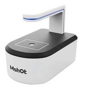
Features: ● Designed for incubator use and long-term imaging ● Phase contrast and fluorescence observation ● Supports multi-well plates and culture vessels ● Automated time-lapse acquisition ● Stable, repeatable positioning
Specifications: ● Model: MCS11 ● Brightfield Observation: ➔ Transmitted phase-contrast observation, working distance: 70 mm ➔ Long-life low-phototoxicity LED: 625 nm ● Fluorescence Observation: None ● Objective Lenses: Single objective system; optional magnifications: 4× / 10× / 20× / 40× ● Stage: Flat specimen stage, manual movement ● Dimensions (W × L × H): 143 × 232 × 182 mm ● Z-axis: Motorized focusing, auto-focus ● Camera: 5.0 MP brightfield camera ● Software Features: C/S architecture with remote control support; includes light control, camera control, image capture, video recording, time-lapse imaging, cell counting, cell confluence analysis, wound healing assay, Web-based control, and email alerts ● Data & Power Interface: USB data cable + DC power cable ● Operating Environment: 5–42 °C, 5–95% RH (non-condensing)
|
| MCS22 | 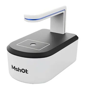
Features: ● Designed for incubator use and long-term imaging ● Phase contrast and fluorescence observation ● Supports multi-well plates and culture vessels ● Automated time-lapse acquisition ● Stable, repeatable positioning
Specifications: ● Model: MCS22 ● Brightfield Observation: Transmitted phase contrast observation, working distance: 70mm Long-life low phototoxicity LED light source: 625nm ● Fluorescence Observation: Blue (B): 470/40nm; Green (G): 570/40nm Three optional fluorescence channels, wavelengths customizable ● Objective Lenses: Dual objectives with electric turret switching Optional magnifications: 4x / 10x / 20x ● Stage: Flat specimen stage, manual movement ● Dimensions (W × L × H): 214 × 261 × 182 mm ● Z-Axis Focus: Electric focusing, autofocus ● Camera: High-speed high-sensitivity fluorescence camera 5.0 megapixels; Sensor size: 2/3″; Frame rate: 40fps ● Software: C/S architecture with remote control support, including functions for: Light control, camera control, image capture, video recording, time-lapse photography, cell counting, cell confluence analysis, scratch assay, Web-based control, and email alerts ● Data & Power Interface: USB data cable + DC power cable ● Operating Environment: Temperature: 5–42°C; Humidity: 5–95% RH (non-condensing)
|
Untuk pertanyaan produk dan stock lebih lanjut Bapak/Ibu dapat menghubungi kontak yang tertera pada laman website Indogen berikut. Terima kasih.
Referensi: